








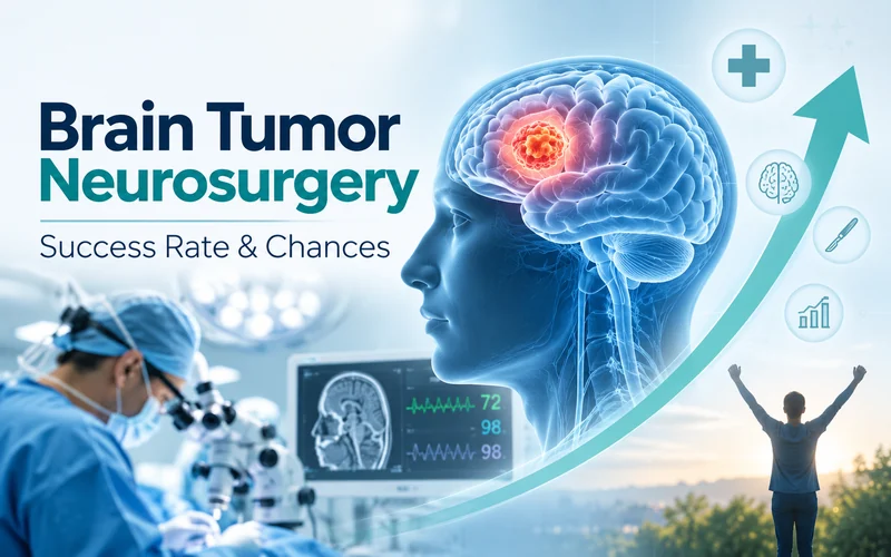
“What’s the success rate?” is the first question almost every patient asks after a brain tumor diagnosis, and it’s also the hardest to answer with one number. Brain tumor surgery outcomes vary enormously depending on tumor type, location, and grade, and understanding these differences matters more than any single statistic quoted in isolation.
There is no single brain tumor surgery success rate. Benign tumors like meningiomas have 85 to 95% survival with complete surgical removal. Low-grade gliomas see 70 to 90% survival. Malignant tumors like glioblastoma are measured differently; according to a neurosurgeon in Delhi, success means maximal safe tumor removal to extend life and preserve function, not a percentage cure rate.
Why “Success Rate” Isn’t a Single Number
Success in brain tumor surgery is defined differently depending on what kind of tumor you actually have. For a benign tumor, success often means complete removal and a return to normal life. For an aggressive malignant tumor, success is measured as “maximal safe resection”, removing as much tumor as possible without damaging critical brain function, since complete cure isn’t always realistic even with modern techniques.
Brain Tumor Types and Their Surgical Outlook
Your specific tumor type is the single biggest driver of your outlook.
| Tumor Type | Typical Outlook |
| Meningioma (benign) | Complete removal success: 90–95% |
| Pituitary adenoma (benign) | Complete removal success: 90–95% |
| Low-grade glioma (Grade 1–2) | Favourable survival: 70–90% |
| Oligodendroglioma | 69–90% depending on age at diagnosis |
| Glioblastoma (Grade 4) | Median survival 12–18 months; maximal safe resection is the goal |
These figures come from published international outcome data and vary with individual factors; they’re a starting reference point for informed conversations with your surgeon, not a personal prediction.
What Actually Determines Your Individual Chances
Beyond tumor type, several factors shape your specific outlook.
| Factor | Why It Matters |
| Tumor location | Areas near motor, speech, or deep midline structures are harder to fully resect |
| Tumor grade | Higher-grade tumors are more likely to recur even after surgery |
| Patient age | Younger patients generally tolerate surgery and recover function better |
| Overall performance status | Baseline strength and independence affects surgical candidacy and recovery |
| Extent of resection achievable | How much tumor can safely be removed directly affects long-term outcome |
Extent of Resection: The One Factor Surgeons Can Actually Control
Extent of resection (EOR), how much of the tumor is actually removed, is widely considered the most significant factor a surgeon can influence to improve long-term outcomes. This is precisely why the surgical approach and the surgeon’s experience with your specific tumor type and location matters so much, tumors near the motor strip, skull base, or deep midline structures are technically harder to fully resect regardless of how advanced the equipment is.
Modern Techniques That Improve Outcomes
- Neuronavigation systems act like GPS for the brain, guiding the surgeon with real-time, image-based precision during the operation
- Intraoperative MRI allows surgeons to scan the brain mid-surgery, confirming how much tumor has actually been removed before closing
- Awake craniotomy, keeping the patient conscious during specific parts of the surgery, lets surgeons test language and motor function in real time when the tumor sits near critical areas
- Neuromonitoring tracks nerve and brain function continuously throughout the procedure, reducing the risk of new neurological deficits
What Happens During Brain Tumor Surgery
Most brain tumor surgery follows a craniotomy approach: a section of skull is temporarily removed to access the tumor, which is then removed using microsurgical techniques guided by neuronavigation. Depending on tumor type and location, this may be combined with awake mapping or intraoperative imaging. Once the procedure is complete, the bone is repositioned and secured, and the patient moves to intensive monitoring.
Recovery and Follow-Up
Hospital stays typically range from a few days to two weeks depending on tumor complexity and any complications. Physical, speech, or occupational therapy may follow if the tumor or surgery affected specific brain functions. For malignant tumors, radiation or chemotherapy often follows surgery as part of a combined treatment plan, and regular imaging follow-up continues for years to monitor for recurrence, particularly with tumors known to regrow even after apparently complete removal.
When to See a Neurosurgeon in Delhi
Persistent headaches that worsen over time, new seizures, unexplained weakness or numbness, vision or speech changes, or personality shifts noticed by family members all warrant prompt neurological evaluation. Early diagnosis from a neurosurgeon in Delhi genuinely changes surgical options, smaller, earlier-stage tumors are generally more amenable to complete or near-complete removal than those discovered after significant growth.
Brain tumor surgery success isn’t one number, it’s a picture built from tumor type, location, grade, and how much can be safely removed. Understanding your specific situation, rather than an average statistic, is what actually helps you make informed decisions. Primus Hospital’s neuro sciences team in Delhi offers the imaging, surgical expertise, and follow-up care needed to give you a realistic, individualised picture of your own chances.
Primus Hospital has a dedicated centre for brain and spine surgery, backed by 250 beds, 18 ICU beds, and 6 HDU beds, and advanced imaging. The hospital’s neurosurgery team includes specialists with over two decades of experience specifically in brain and spine tumor surgery, and membership with the American Association of Neurosurgeons. As a leading neurosurgery hospital in Delhi, Primus combines this surgical experience with round-the-clock neurology and critical care support for both routine and complex cases.
Learn more or book a consultation at primushospital.com.